

https://www.cellstrains.co...
Get an EXTRA 20% OFF your hosting plan!
Hostinger's got a HOT deal! Get an EXTRA 20% OFF your hosting! Don't miss out!

Cell imaging systems allow scientists to observe cells and their activities in real-time without direct human intervention. These automated systems use advanced microscopy techniques and image capturing capabilities to gain insights into cellular processes.
One of the most widely used microscopy techniques in cell imaging is fluorescence microscopy. It utilizes fluorescent probes or fluorescent proteins to label specific parts of cells or target molecules. When exposed to light of a specific wavelength, these fluorescently tagged elements emit light of a longer wavelength that can be detected by the microscope. This non-invasive approach enables observation of live cells and dynamic cellular events over long periods.
Get More Insights On Automatic Cell Imaging System
https://www.exoltech.us/bl...

Automatic Cell Imaging System: A revolutionary change in medical...
Cell imaging is a crucial element of medical diagnostics and research. Cells contain a wealth of information about the human body and its functions. By analyzing cells under a microscope, scientists and doctors can gain valuable insights. Traditional cell imaging relies on manual...
https://www.exoltech.us/blogs/280491/Automatic-Cell-Imaging-System-A-revolutionary-change-in-medical-diagnosticsRead More: https://www.imarcgroup.com...
Advancements in Cryo-EM Imaging Technology
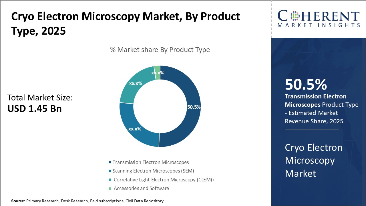
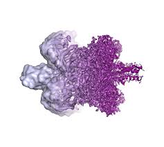
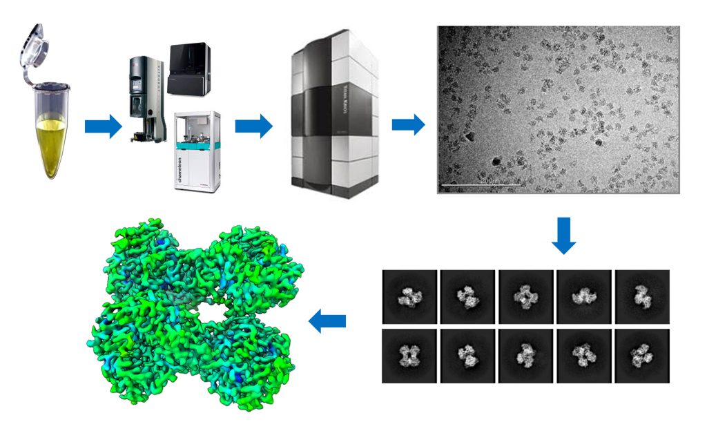
Cryo electron microscopy (Cryo-EM) allows visualization of biological molecules at nearatomic resolution by flash-freezing biological samples and imaging them with electron microscopes. It is being used to image macromolecular complexes such as protein-protein and protein-nucleic acid complexes. Cryo-EM offers significant advantages over X-ray crystallography such as ability to image large complexes, flexibility in sample purity and quantity requirements.
The Global Cryo Electron Microscopy Market is estimated to be valued at USD 1.45 Billion in 2025 and is expected to reach USD 3.18 Billion by 2032, exhibiting a compound annual growth rate (CAGR) of 11.9% from 2025 to 2032.
Cryo Electron Microscopy Market
https://www.coherentmarket...
Get More Insights On Cryo Electron Microscopy Market
https://www.zupyak.com/p/4...
#CryoElectronMicroscopyMarket , #CryoElectronMicroscopyMarketsize , #CryoElectronMicroscopyMarketshare , #CryoElectronMicroscopyMarketApplication , #CryoElectronMicroscopyMarkettrends
Cryo Electron Microscopy Market Size and Forecast, 2025-2032
Cryo Electron Microscopy Market valued at USD 1.45 Billion in 2025 , is anticipated to reaching USD 3.18 Billion by 2032, with a steady CAGR of 11.9% .
https://www.coherentmarketinsights.com/industry-reports/cryo-electron-microscopy-marketCryo electron microscopy (cryo-EM) is a microscopy technique used to obtain highresolution 2D structure images and 3D structures of macromolecules, viruses, and cellular components which are rapidly frozen to cryogenic temperatures. It involves the use of a transmission electron microscope to examine biomolecules that have been frozen in a thin layer of vitrified ice. Cryo-EM allows the observation of biomolecules in a near-native hydrated state without the need for crystallography and can achieve resolutions well beyond the limits of light microscopy.
The growing demand for characterization of macromolecules at near-atomic resolutions without the requirement of crystallization is one of the major factors driving the growth of the cryo EM market.
The Global Cryo Electron Microscopy Market is estimated to be valued at USD 1.45 Billion in 2025 and is expected to reach USD 3.18 Billion by 2032, exhibiting a compound annual growth rate (CAGR) of 11.9% from 2025 to 2032.
Get More Insights On Cryo Electron Microscopy Market
https://www.zupyak.com/p/4...
Cryo Electron Microscopy Market is Growing Rapidly Driven by Technological Advancements | Zupyak
https://www.zupyak.com/p/4565990/t/cryo-electron-microscopy-market-is-growing-rapidly-driven-by-technological-advancements
Life Science Instrumentation Market
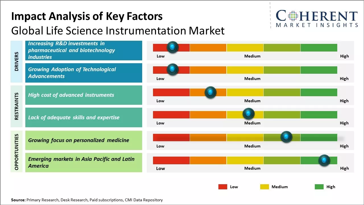
Life science instrumentation encompasses a broad array of analytical devices—such as chromatography systems, mass spectrometers, flow cytometers, DNA sequencers, and advanced microscopy platforms
Life Science Instrumentation Market - https://www.coherentmarket...
Life Science Instrumentation Market Size & Forecast, 2032
Life Science Instrumentation Market is growing with a CAGR of 6.4% in the prediction period and it crosses USD 90.58 Bn by 2032 from USD 58.63 Bn in 2025
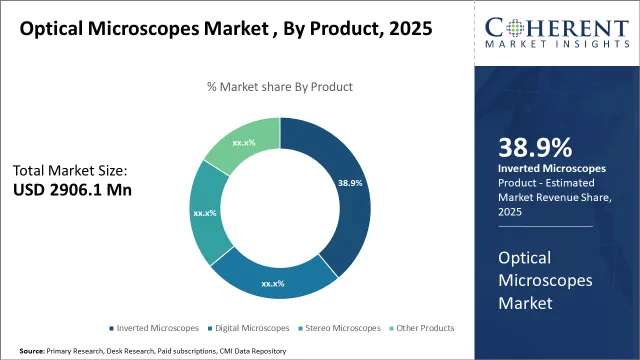
https://www.coherentmarketinsights.com/industry-reports/life-science-instrumentation-marketOptical microscopes remain indispensable instruments in biological research, materials science, and clinical diagnostics due to their highresolution imaging, versatility, and relatively low cost compared with electron microscopy. These instruments employ visible light and glass lenses to magnify samples, enabling brightfield, darkfield, phasecontrast, and fluorescence techniques that reveal cellular structures, surface topography, and livecell dynamics.
Recent advances in optics, digital imaging, and software‐driven automation have expanded the market scope, allowing seamless integration with AI for realtime analysis. Laboratories benefit from faster workflows, improved image clarity, and minimized downtime, driving business growth across pharmaceutical R&D, semiconductor inspection, and academic institutions.
The global optical microscopes market is estimated to be valued at USD 2,906.1 Mn in 2025 and is expected to reach USD 4,266.9 Mn in 2032, exhibiting a compound annual growth rate (CAGR) of 5.64% from 2025 to 2032.
Optical Microscopes Market
https://www.coherentmarket...
Get More Insights On Optical Microscopes Market
https://justpaste.it/g0nfd
Optical Microscopes Market Size, Share & Analysis, 2025-2032
Optical Microscopes Market valuation is estimated to reach USD 2,906.1 Mn in 2025 and is anticipated to grow USD 4,266.9 Mn by 2032 with steady CAGR of 5.64%.
https://www.coherentmarketinsights.com/market-insight/optical-microscopes-market-5606Cryo electron microscopy (cryo-EM or cryo-TEM) is a powerful technique used to determine the structure of biomolecules at near-atomic resolution. In cryo-EM, biological samples such as proteins, nucleic acids, and protein complexes are rapidly frozen in a thin film of vitreous ice and imaged using an electron microscope. Cryo-EM overcomes many of the limitations of X-ray crystallography by not requiring crystallization of samples. This allows structure determination of more heterogeneous and dynamic molecules.
Get More Insights on Cryo Electron Microscopy
https://www.patreon.com/po...
#CryoElectronMicroscopy #StructuralBiology #Biomolecules #Macromolecules #CoherentMarketInsights
The Global Asbestos Testing Market is estimated to be valued at US$ 242.3 Mn in 2025 and is expected to exhibit a CAGR of 5.2% over the forecast period 2025 to 2032.
Asbestos testing involves the systematic analysis of building materials, soil, air, and water samples to detect and quantify asbestos fibers, protecting public health and the environment. Products in this market range from sample collection kits and laboratory reagents to portable fiber analyzers and advanced microscopy systems. These solutions offer high sensitivity, accuracy, and rapid turnaround times, enabling stakeholders to identify potential risks in older structures, industrial sites, and renovation projects. Continuous monitoring and periodic assessments help facility managers and environmental consultants mitigate health hazards, optimize maintenance budgets, and align with evolving regulatory drivers. Furthermore, the integration of digital data reporting platforms enhances transparency across supply chains and supports data-driven decision-making.
Get more insights on, Asbestos Testing Market- https://justpaste.me/PLHo4
#CoherentMarketInsights #AsbestosTesting #AsbestosTestingMarket #AsbestosTestingMarketInsights #BulkMaterialTesting #AirQualityTesting

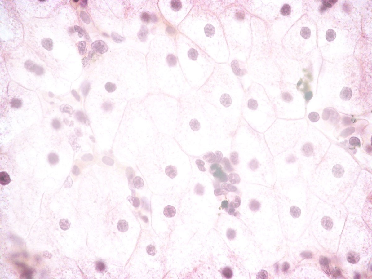
Histology and cytology technologies encompass a suite of diagnostic tools and consumables—ranging from staining reagents, automated slide stainers, microscopy systems, immunohistochemistry kits to molecular probes
The rising Histology and Cytology Market Demand
for minimally invasive diagnostic procedures and the growing prevalence of cancer and chronic diseases present significant market opportunities.
Histology and Cytology Market - https://www.coherentmarket...
#AIPoweredDigitalPathology
#PrecisionCancerDiagnostics
#HistologyAndCytologyMarket
#HistologyAndCytologyMarketTrends
#HistologyAndCytologyMarketDemand
#CoherentMarketInsights
The Cryo-Electron Microscopy (Cryo-EM) Structure Analysis Services Market has emerged as a game-changer in the field of structural biology, offering unprecedented insights into the intricate world of biomolecules. This cutting-edge technology has revolutionized the way researchers study.
Cryo-EM has rapidly gained prominence in recent years due to its ability to visualize biological structures at near-atomic resolution without the need for crystallization, which has been a major bottleneck in traditional X-ray crystallography.
Get more insights on, Cryo-EM Structure Analysis Services Market- https://www.patreon.com/po...
#CoherentMarketInsights #Cryo -EMStructureAnalysisServicesMarket #Cryo -EMStructureAnalysisServices #Cryo -EMStructureAnalysisServicesMarketInsights #ElectronCrystallography
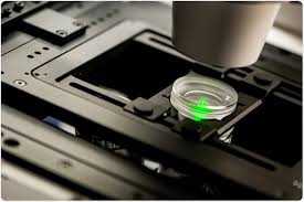
The Global Two-Photon Microscopy Market is estimated to be valued at US$ 1.35 Bn in 2025 and is expected to exhibit a CAGR of 8.7% over the forecast period 2025 to 2032.
Two-photon microscopy is an advanced fluorescence imaging technique that enables deep tissue visualization with minimal phototoxicity, making it indispensable for applications in neurobiology, cancer research, and developmental biology. Employing near-infrared lasers, this technology uses simultaneous absorption of two photons to excite fluorophores, providing high-resolution, three-dimensional images up to one millimeter beneath the specimen surface. Key components include ultrafast pulsed lasers, high-sensitivity detectors, precision scanning optics, and specialized high numerical aperture objectives.
Get more insights on, Two-Photon Microscopy Market- https://www.patreon.com/po...
#CoherentMarketInsights #Two -PhotonMicroscopyMarket #Two -PhotonMicroscopyMarket #Two -PhotonMicroscopyMarket Insihts #microscopes
In recent years, the field of pathology has witnessed a significant transformation with the advent of digital slide scanners. These innovative devices have revolutionized the way pathologists analyze and interpret tissue samples, offering numerous benefits over traditional microscopy methods.
Digital slide scanners are sophisticated imaging devices that capture high-resolution digital images of microscope slides. These scanners employ advanced optical systems, including high-quality lenses and cameras, to digitize the entire slide at multiple magnifications.
Get more insights on, Digital Slide Scanners- https://www.patreon.com/po...
#CoherentMarketInsights #DigitalPathology #Telepathology #LabDiagnostics #PrecisionMedicine

The Global Virus Safety Testing Services Market is estimated to be valued at US$ 682.4 Mn in 2025 and is expected to exhibit a CAGR of 16.50% over the forecast period 2025 to 2032.
The Virus Safety Testing Services Market encompasses a comprehensive suite of assays, quality control measures, and validation protocols designed to detect viral contaminants in biopharmaceutical products, vaccines, and gene therapies. These services include in vivo and in vitro tests such as adventitious agent screening, PCR-based detection, cell culture-based assays, and electron microscopy. Advantages of outsourcing virus safety testing include access to specialized expertise, state-of-the-art laboratories, and scalable testing capacities that support rapid product development timelines. Biopharmaceutical companies increasingly seek third-party testing to mitigate risks, ensure compliance with cGMP standards, and streamline market entry. The market’s growing emphasis on product purity and patient safety drives demand for advanced testing protocols, bolstering market growth.
Get more insights on, Virus Safety Testing Services Market- https://justpaste.me/PLUy6
#CoherentMarketInsights #VirusSafetyTestingServicesMarket #VirusSafetyTestingServices #VirusSafetyTestingServicesMarketInsights #ViralDetectionTesting #ViralClearanceTesting

Tamil Nadu boasts a network of highly skilled gemmologists renowned for their expertise in gemstone identification, valuation, and certification. Certified professionals in cities like Chennai, Coimbatore, and Madurai leverage advanced technologies such as spectroscopy and microscopy to ensure accurate gemstone analysis. Specializing in diamond grading, synthetic gem detection, and custom jewellery appraisal, these experts adhere to global gemological standards. Many hold certifications from internationally recognized institutions and offer tailored services for antique jewellery restoration, investment-grade gemstones, and ethical sourcing guidance.
For a comprehensive PDF guide on Tamil Nadu’s top gemologists, optimize your content with keywords like "certified gemologist Tamil Nadu," "gemstone testing services," and "professional gem certification." Use meta descriptions like "Explore trusted gemology experts in Tamil Nadu" to enhance search visibility.
Discover Best Gemologist in Tamilnadu offering certified gemstone testing, diamond grading, and expert jewellery appraisal services. Download now
https://navagrahaonline.co...

Expert Gemologist Services in Kerala & Tamil Nadu | Navagraha Online
Looking for trusted gemologist services in Kerala and Tamil Nadu? Navagraha Online offers professional gemstone consultation, Vedic astrology-based gems, and personalized recommendations to enhance your well-being and fortune. Get expert guidance today!
https://navagrahaonline.com/gemologist-services/